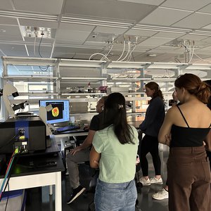
Microscopy Class Demos State of the Art Microscopes
BIO400/600 students demo some of the best microscopes in the market.

The Blatt BioImaging Center is a light microscopy core facility that focuses on live cell and enhanced resolution imaging of diverse model organisms ranging from cells to organoids and whole organisms. Located on the 2nd and 3rd floors in the Biology Department in the Life Sciences Complex (Rooms 249, 259, 359 and 318) the imaging center houses state-of-the art confocal microscopes and high-resolution bright field imaging systems. The resources and services of the Blatt BioImaging Center are open to biomedical scientists (students, staff and faculty) at Syracuse University and external investigators at both academic institutions and commercial enterprises in the Upstate New York area.
Banner image courtesy of L. Rathbun.
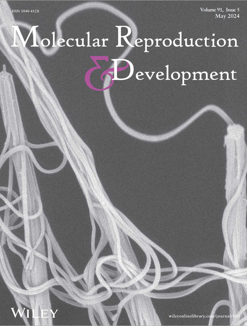
Description: Scanning Electron Microscope image of spermatostyle morphology in Dineutus assimilis whirligig beetles. Multiple spermatostyles with numerous attached sperm are shown. Spermatostyles were isolated from male seminal vesicles. Read more:https://doi.org/10.1002/mrd.23745
Image Credit: Antonio Gomez, Yasir Ahmed-Braimah, Scott Pitnick, Steve Dorus.
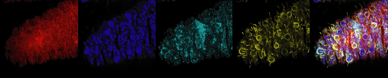
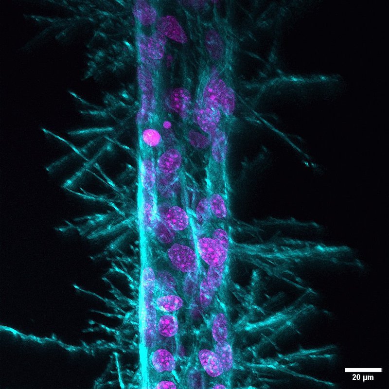
Close up confocal micrograph of a Hydra tentacle. The image highlights the cytoskeleton (red), DNA (blue), centrosomes (cyan) and microtubules (yellow).
Courtesy A. Aljiboury"
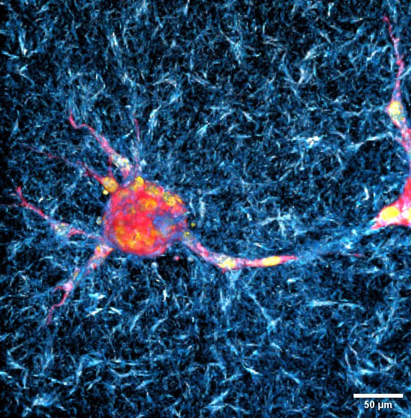
Zeiss LSM 980 Confocal Image of a Wild-type mouse embryonic fibroblast spheroids invasion in 3D Collagen Gel. Concentration: 1.5mg/ml Collagen I Rat Tail. Scale bar is provided. Color: Red/Magenta: Actin, Yellow: Nucleus, Blue/White: Collagen Fibril
Courtesy of Minh-Tri Ho Thanh
A collaboration between Blatt BioImaging center users, the Physics Department’s Patteson lab, and the Department of Biomedical and Chemical Engineering’s Soman lab produces cool microscopy images!
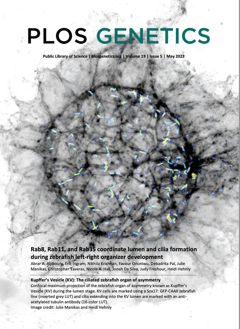
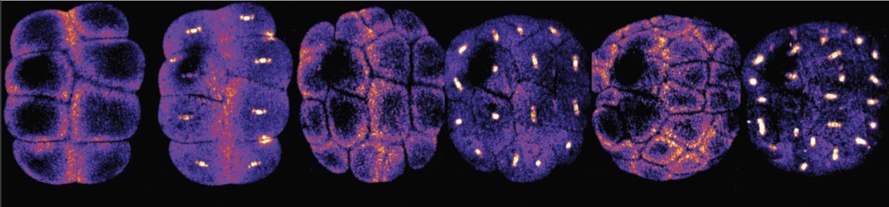
Title: Rab8, Rab11, and Rab35 coordinate lumen and cilia formation during zebrafish left-right organizer development
Authors: Abrar A. Aljiboury, Eric Ingram, Nikhila Krishhan, Favour Ononiwu, Debadrita Pal, Julie Manikas, Christopher Taveras, Nicole A. Hall, Jonah Da Silva, Judy Freshour, Heidi Hehnly
Description: Kupffer's Vesicle (KV): The ciliated zebrafish organ of asymmetry Confocal maximum projection of the zebrafish organ of asymmetry known as Kupffer's Vesicle (KV) during the lumen stage. KV cells are marked using a Sox17: GFP-CAAX zebrafish line (inverted grey LUT) and cilia extending into the KV lumen are marked with an anti-acetylated tubulin antibody (16-color LUT).
Image credit: Julie Manikas and Heidi Hehnly
BIO400/600 students demo some of the best microscopes in the market.
Bioimaging tools are used to create BioArt and pushing the limits of visual arts.
The SU BioImaging center invites you to join us for a symposium introducing analytical resources on the Hill and FREE Microscope demo opportunities.