
How Physics Could Improve IVF Success
Key Takeaways:
- Chromosome Errors: Human egg cells often fail to properly segregate their chromosomes, with consequences including miscarriage and failed IVF cycles.
- Imaging Innovation: Non-invasive imaging techniques being developed and tested by a Syracuse University physicist measure the stability of a structure known as the spindle to assess egg quality.
- Link to Fertility Outcomes: This approach could help clinics identify the eggs most likely to succeed before implantation and tailor growth conditions individually, raising IVF success rates and lowering costs for patients.
At the intersection of physics and human reproduction, Colm Kelleher is helping answer one of fertility medicine's most persistent questions: why do some human eggs fail to divide their chromosomes correctly? Errors in this process can mean a pregnancy that ends before it begins, or an IVF cycle that doesn't succeed. Kelleher, an assistant professor of physics in the College of Arts and Sciences and a member of the University's BioInspired Institute, is trained as a biophysicist, but his work is poised to reshape how scientists assess egg and embryo quality.
A Cellular Machine Built to Move Chromosomes
As egg cells develop, they must physically move their chromosomes into position so that, if fertilized, the resulting embryo receives the correct number of chromosomes. To do this, egg cells build a specialized structure called the spindle, which attaches to chromosomes and exerts the forces needed to separate them properly.
"This research will develop a promising method to measure the spindle, and investigate whether the resulting data can tell us about the potential of individual egg cells to develop successfully," Kelleher says.

Colm Kelleher
Chromosome segregation errors are a major cause of embryonic aneuploidy (a condition in which an embryo has too many or too few chromosomes), miscarriage and failed embryo development in in vitro fertilization (IVF). Kelleher's project, "Quantitative Links Between Cellular Energy Availability, Spindle Function and Chromosome Segregation in Human Oocytes," funded by a grant from the American Society for Reproductive Medicine (ASRM), aims to close a knowledge gap and get to the root cause of those segregation errors.
Partnering with Fertility Clinics
The Kelleher lab is developing non-invasive imaging techniques to measure spindle structure in living oocytes (immature egg cells). The technique relies on polarized light microscopy, which uses the way light passes through the spindle to reveal details about its structure without damaging the egg. This is similar to how X-rays allow doctors see bone structure without harming the patient.
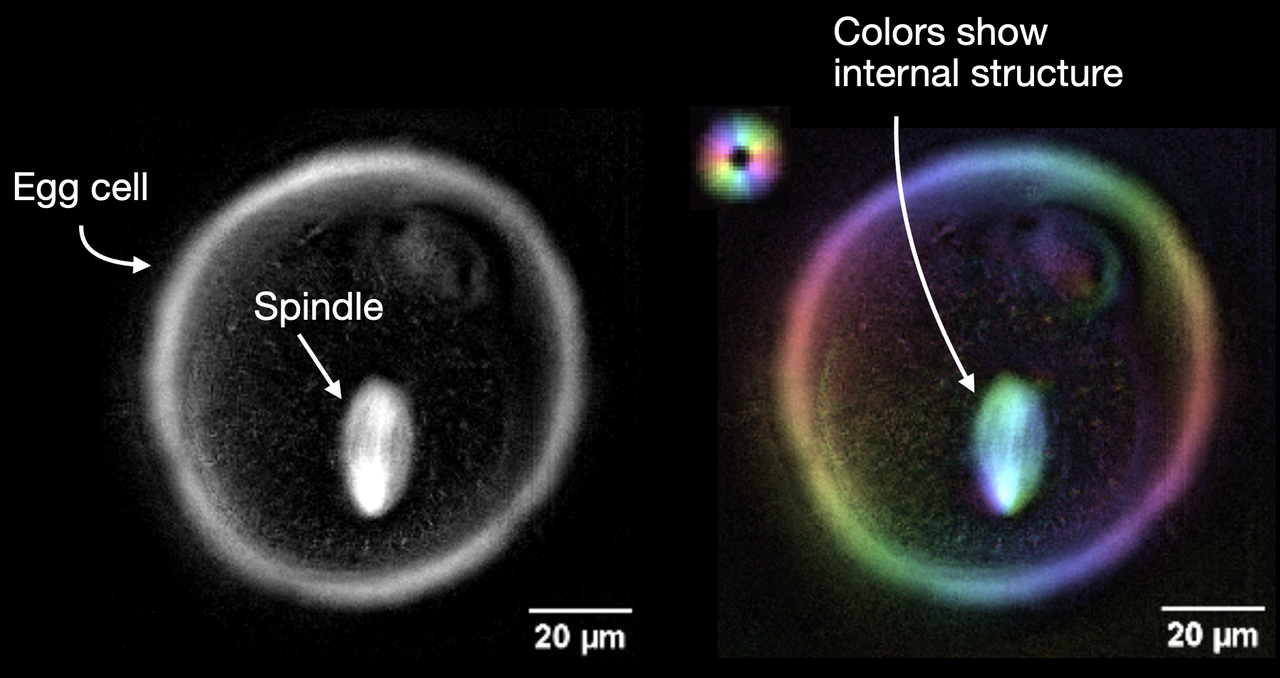
Polarization microscopy of spindles isn't new; it's already used in IVF clinics. "Our work builds on this by using computational image analysis and physics-based modeling to extract properties of the spindle that could not be measured previously," Kelleher says.
Prior work showed the technique can measure spindle properties in mouse egg cells, but mice are not a perfect stand-in for humans. Chromosome segregation fails far more often in human eggs, so testing the method in human eggs required a research partner.
"It is essential to directly test these methods in human eggs, which can only be obtained by partnering with IVF clinics such as CNY Fertility and Boston IVF," Kelleher says. The eggs donated by those clinics and used in the research are ones that would not have been used for IVF treatment because they might be unsuitable for fertilization, he notes.
Because the imaging technique is non-invasive and doesn't damage the egg, Kelleher envisions it eventually being used to directly assess individual eggs under consideration for fertilization in IVF clinics. Looking further ahead, he says similar methods could even be used to tailor growth conditions for each individual egg or embryo, improving the odds that at least one develops successfully. Together, those applications could raise IVF success rates and lower treatment costs.
What the Grant Makes Possible
As Syracuse's first recipient of an ASRM grant, Kelleher says the award carries meaning beyond the funding itself. "It will fully fund a graduate student and a postdoc for several years, which will allow us to make progress much faster," he says. "I am somewhat of an outsider in the IVF field, so it is encouraging that the reproductive medicine community finds our work of interest, and great for my industry partners at CNY Fertility to see this external validation."
The lab has nearly finished setting up its microscopes and has already imaged spindles in mouse egg cells for practice. It recently received its first shipments of frozen human egg cells and expects to begin imaging them by the end of summer.
The ASRM Research Institute serves as a catalyst for groundbreaking discoveries in areas often overlooked by traditional funding sources. Kelleher's award reflects that mission, bringing a physicist's toolkit to bear on one of reproductive medicine's most consequential open questions.
Published: July 28, 2026
Media Contact: asnews@syr.edu